Michael Hooker Microscopy Facility (MHMF.ORG)
![]()
|
|
Michael Hooker Microscopy Facility (MHMF.ORG) |
|
C-Imaging - SimplePCI - Cell Counting
Index
0. Introduction
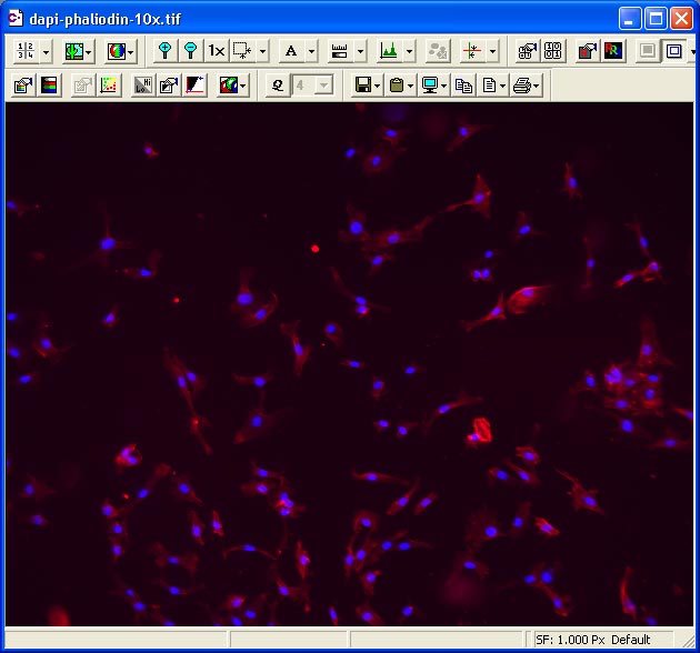
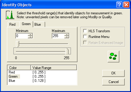
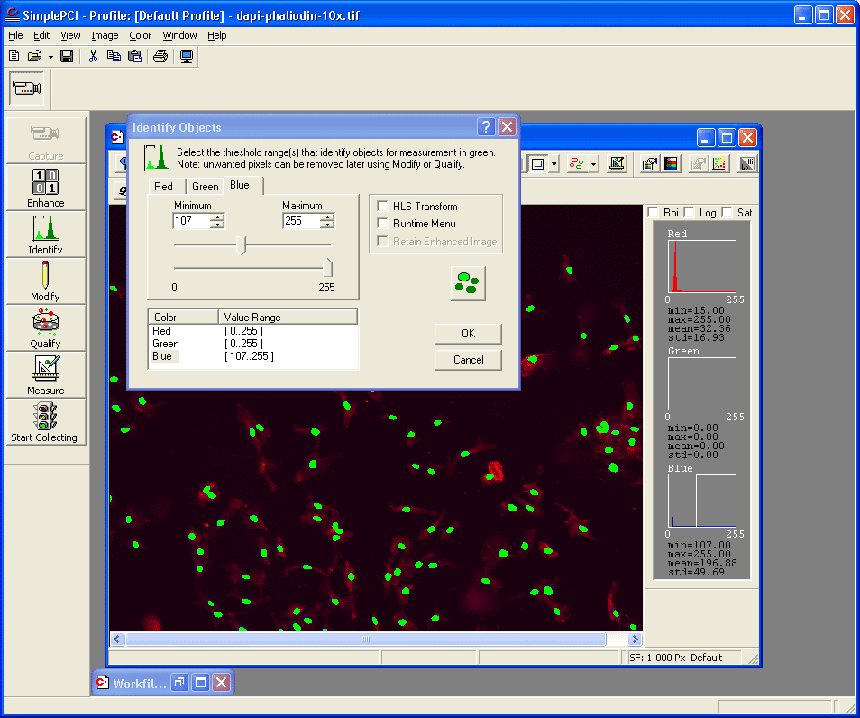
An effective way to count cells is to fluorescently label them with the DNA binding dye DAPI. This will effectively outline the nuclei, which are inherently separated from each other by cytoplasm. This separation enhances automatic counting. Below are outlined the typical steps used to count cells in images acquired as tiff images or .cxd images. DAPI is effective for fixed permeabilized cells. For live cells several suitable dyes exist. e.g. Hoechst 33342, Draq-5 or Sytox Green.
Care should be exercised to obtain the best quality image possible. Image exposure should be set to give the best contrast possible. i.e. Brightest nuclei relative to the general back ground. It may be advantageous to carry out back ground subtraction. Also staining should be carefully done without over staining with too high a concentration of DAPI, or leaving DAPI in the mounting media, both of which will yield significant background fluorescence, which will reduce contrast. Overexposure of the nuclei is not necessarily detrimental unless flaring of the image spreads to such an extent that the imaged nuclei touch each other. Also the fluorescence light path field aperture and aperture should be fully opened in order to give the most even illumination across the field of view.
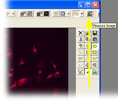
1. Counting from already acquired image(s)


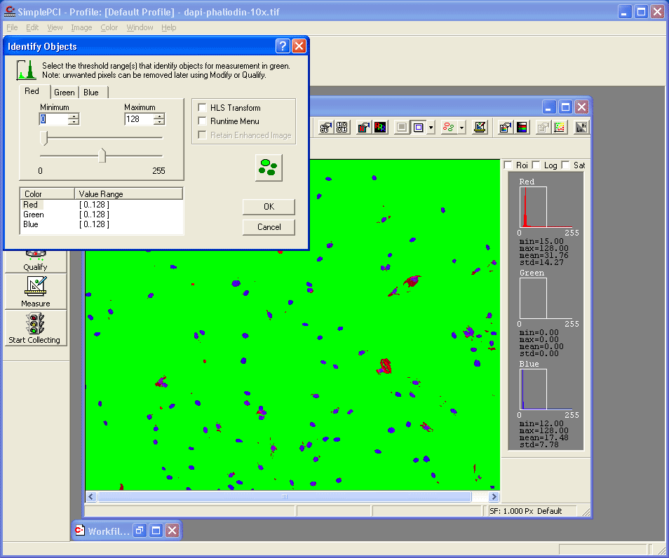
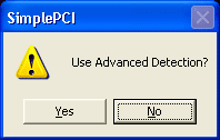
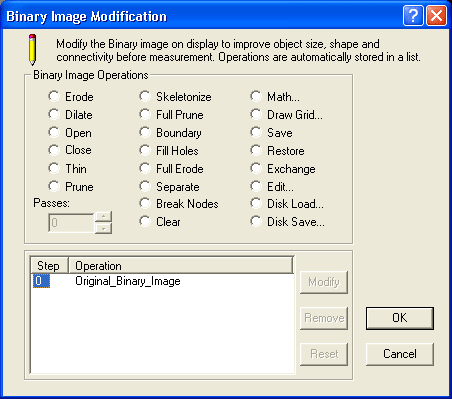
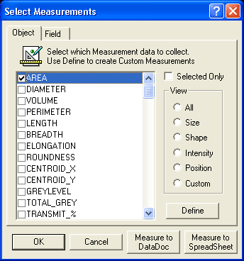
- Note: Advanced Detection has many capabilities, e.g. Separation of touching objects
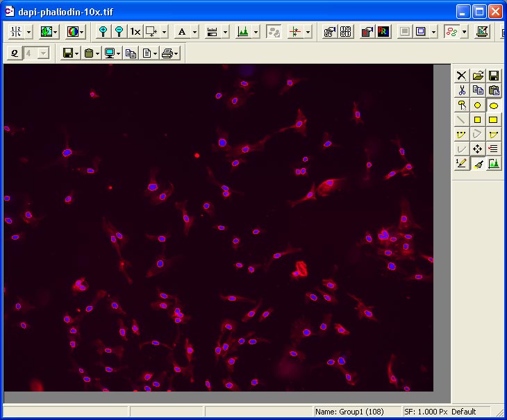
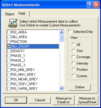
- Or select to measure, say, "Object Area" (Although not going to measure area need to select at least one parameter, so that counting is performed)
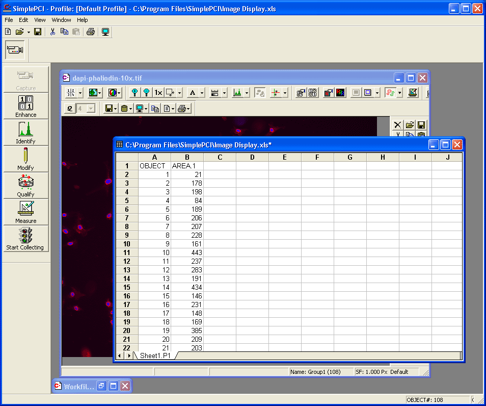
- Or press "Measure to SpreadSheet" in order to retain measurements in a table.
|
|
|
Copyright 2001-2015 Dr. M. Chua, School of Medicine, University of North Carolina, Chapel Hill, NC 27599 |
| Go Back | Booking Resources |
Questions/comments/problems: Michael Chua |
|
|
Last Updated: 2014-07-24 |